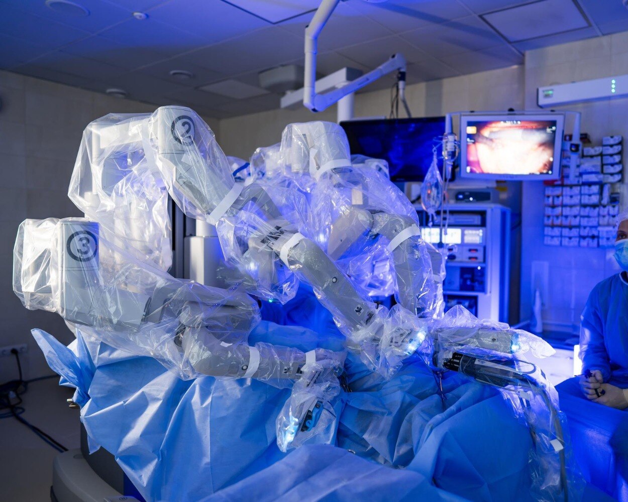
Precision in cancer surgery begins long before the robot enters the operating room. You need to understand what the system actually contributes, how the team verifies cancer clearance, and which hospital capabilities matter when the operation is complex.
Key takeaways
- A surgeon, not the robot, controls every instrument movement in real time.
- Review imaging, biopsy results and surgical goals before consenting to robotic cancer surgery.
- Ask how the team verifies positioning, instruments, pathology and recovery after surgery.
- Choose a hospital with relevant cancer expertise, trained staff and clear follow-up plans.
What “precision” means when a surgeon uses a robot
Precision in robotic cancer surgery means controlled movement and clearer visualisation, not an autonomous operation. A surgeon operates a surgeon-controlled robotic system in real time. It translates hand movements into instrument movements, filters tremor and provides a stable, three-dimensional magnified view.
Wristed instruments can bend and rotate inside the body, helping with dissection and suturing in confined spaces such as the deep pelvis.
| Option | Technical advantage | What it does not guarantee |
|---|---|---|
| Robotic surgery | Magnified 3D view and wristed instruments | Clear margins, complete cancer removal or fewer complications |
| Laparoscopic surgery | Small incisions and direct surgeon control | Easier access to every confined space |
| Open surgery | Broad access and direct tactile handling | A less demanding recovery |
The robot does not identify cancer, choose a dissection plane or decide whether a margin is clear. It cannot guarantee adequate lymph-node assessment, preservation of nerves or blood vessels, or a longer survival.
For colon cancer, examination of at least 12 regional lymph nodes is a commonly used quality benchmark, but the final count also depends on the specimen and pathology processing.
Deep pelvic anatomy can make robotic access technically useful, but technical ease is not proof of a better cancer outcome. The ROLARR trial found no statistically significant overall reduction in conversion to open surgery compared with laparoscopy for rectal cancer.
Conversion remains a safety decision when bleeding, poor visibility, dense adhesions or unexpected invasion threatens safe, cancer-complete surgery.
How planning before surgery creates a precise operation
The robot is suitable only when the preoperative evidence shows that it can remove the tumour with an adequate margin, assess the expected lymph nodes and avoid unacceptable injury. It cannot compensate for incomplete diagnosis or staging.
1. Confirm the diagnosis. A biopsy and pathology review establish the tumour type, grade and features that influence treatment. Ask whether the pathology has been reviewed by a specialist team, especially when the operation depends on distinguishing invasive cancer from a less aggressive lesion.
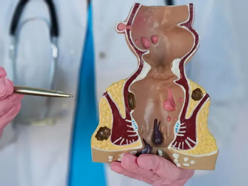
2. Map the disease. CT scan and MRI interpretation show the tumour’s location, size and possible spread. For rectal cancer, high-quality pelvic MRI assesses the mesorectal fascia, sphincter complex and threatened margins; those findings can change chemotherapy, radiation and the planned operation.
3. Complete cancer staging. The team combines scans, examination and pathology to determine whether disease is confined to the primary site, has reached lymph nodes, or has spread elsewhere. Lymph nodes and nearby vessels assessment helps define the dissection required and whether a robotic approach can achieve cancer clearance safely.
4. Review the case together. Surgeons, medical and radiation oncologists, radiologists, pathologists and specialist nurses may decide the treatment sequence as a group. The plan may favour chemotherapy or radiation before surgery, an open operation, or conversion during surgery if bleeding, adhesions or unexpected invasion threaten a sound resection.
For colon cancer, examination of at least 12 regional lymph nodes is a common quality benchmark, although the specimen and pathology processing affect the count. Final margins and microscopic invasion are confirmed by pathology, not by the robot’s view.
How the operating-room team protects precision during the procedure
Precision depends on a controlled sequence of checks, not on the robot alone. The surgeon directs every instrument while the operating-room team verifies anatomy, protects critical structures and responds quickly when conditions change.
1. The team confirms port placement before dissection begins. Correct spacing gives the instruments a clear working angle and prevents collisions, restricted movement or unsafe traction.
2. The surgeon identifies the tumour and nearby structures against the preoperative scans. Careful dissection preserves nerves, ureters, bowel and major vessels when cancer clearance allows; nerve and blood-vessel protection cannot justify leaving involved tissue behind.
3. The team maintains bleeding control through measured dissection, sealing or clipping vessels, and prompt suction. Anesthesia tracks blood pressure and blood loss, while access to blood-bank support and intensive care strengthens the response to a major bleed.
4. The surgeon reassesses the planned operation if visibility is poor, adhesions are dense, bleeding remains uncontrolled or the tumour invades an unexpected structure. Converting to open surgery can be the safer way to protect the patient and achieve an oncologically sound resection.
5. Specimen removal includes checking that the intended organ, tumour-bearing segment and required lymphatic tissue are intact. The team labels and orients the specimen for pathology; a visually complete operation does not prove that microscopic margins are clear.
Final pathology determines margin status, tumour stage, lymphovascular or perineural invasion and lymph-node findings. If a margin is uncertain during surgery, selected hospitals use frozen-section pathology, but the complete cancer assessment usually follows permanent tissue processing.
How to decide whether robotic assistance and a hospital are suitable
Choose the operation and team, not the robot alone. Selected prostate, colorectal, uterine, kidney, stomach, esophageal and thoracic procedures may suit robotic assistance when the tumour’s location, size, spread, prior operations and access to surrounding structures make controlled dissection practical.
| Option | Strength | When it may be preferable |
|---|---|---|
| Robotic surgery | Three-dimensional magnified vision and wristed instruments | Confined spaces or intricate suturing, with a team experienced in that exact cancer operation |
| Laparoscopic surgery | Small incisions and established minimally invasive technique | When the surgeon can achieve the same cancer clearance without robotic assistance |
| Open surgery | Direct access and broad exposure | Large or invasive tumours, dense adhesions, major bleeding, or uncertain anatomy |
Robotic versus laparoscopic versus open surgery is not a ranking from newest to oldest. If visualization is poor, bleeding cannot be controlled, adhesions prevent safe progress, or the surgeon doubts an oncologically sound resection, conversion to open surgery protects the patient; avoiding it at all costs can leave unsafe surgery or inadequate margins.
Assess capability by asking for the surgeon’s case volume with your specific procedure, conversion and complication rates, and who provides backup. Check whether the hospital has a consistent operating team, anesthesia and positioning protocols, blood-bank and intensive-care access, and pathology that reports margins, lymph nodes, stage, lymphovascular and perineural invasion.
Sai Vedant Multi-Speciality Hospital should be assessed using these operating and pathology details, not the presence of a robotic platform alone. The robotic platform and surgeon experience must match the operation.
Related services
Robotic Surgeries Robotic Surgery at Sai Vedant Multispeciality Hospital, Pune Sai Vedant Multispeciality Hospital offers advanced robotic surgery solutions designed... View service → |
Cancer Surgeries Cancer Surgery at Sai Vedant Multispeciality Hospital, Pune Sai Vedant Multispeciality Hospital provides advanced cancer surgery services for the... View service → |
How pathology and recovery show what precision achieved
A cancer operation is judged by the final pathology report, not by a clear-looking surgical field. The report records surgical margins, tumour stage, lymphovascular or perineural invasion, and lymph-node assessment.
For colon cancer, examination of at least 12 regional lymph nodes is a common quality benchmark, but the count also depends on the specimen, pathology processing and tumour biology.
| Finding | What it establishes | What it does not establish |
|---|---|---|
| Negative surgical margins | No cancer reaches the examined cut edge | That microscopic disease is absent elsewhere |
| Lymph-node assessment | Whether sampled nodes contain cancer and helps determine stage | That unsampled nodes are disease-free |
| Visually complete removal | The intended specimen was removed | Microscopic clearance or long-term cure |
Recovery should follow a defined plan rather than rely on the robot’s advertised benefits. An Enhanced Recovery After Surgery pathway can include limited fasting, opioid-sparing pain relief, early walking and early oral intake; it can reduce complications and shorten admission after major colorectal surgery, but it does not make high-risk surgery low-risk.
Ask the hospital:
- How many millimetres was the closest surgical margin?
- How many lymph nodes were examined, and how many contained cancer?
- Who reviewed the final pathology report, and will you receive it?
- What are the centre’s conversion-to-open-surgery, complication, readmission and reoperation rates for this operation?
- Which recovery milestones are expected, and what happens if you miss them?
- Which findings remain uncertain after pathology, and could they change chemotherapy or radiation treatment?
Frequently asked questions
What does precision mean in robotic cancer surgery?
Precision means surgeon-controlled instrument movement, tremor filtration and a stable, magnified three-dimensional view; the robot does not operate autonomously.
How does planning before surgery improve precision?
The team reviews imaging, biopsy findings, the tumour’s location and the surgical goal to plan access, positioning and possible alternatives.
How does the operating-room team protect precision during surgery?
The team confirms the patient’s position, equipment settings, instrument readiness and communication protocols before and during the procedure.
How can you decide whether robotic surgery and a hospital are suitable?
Ask about the surgeon’s experience with your cancer and procedure, the hospital’s equipment and team, alternatives, risks, conversion plans and follow-up care.
How do pathology and recovery show what precision achieved?
Pathology assesses the removed tissue and margins, while recovery monitoring shows whether treatment goals were met without avoidable complications.